
What is the lens?
The lens is a large transparent structure within the eye lying just behind the black part of the eye (the pupil). The lens helps to focus light onto the back of the eye. It is normally held in place by tiny threads all around its edge.
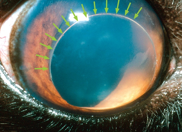
A dislocated (luxated) lens in the front chamber of the eye. The arrows mark the edge of the lens
What happens in lens luxation?
In patients suffering from lens luxation (dislocation), the lens shifts out of position and moves either into the front or into the back of the eye. If the lens becomes trapped in the front of the eye, it can cause water-logging of the window of the eye (seen as blueing of the front of the eye) and an increase in pressure within the eye (glaucoma). If it falls into the back of the eye, the lens can damage the sensitive tissue at the back of the eye (the retina) and cause it to loosen (retinal detachment). This can also lead to glaucoma. In both cases, the eye is usually painful, and blindness develops unless early appropriate treatment is given.
What causes lens luxation?
Many cases of lens luxation are hereditary and caused by a weakness in the threads holding the lens in place. This condition is called primary lens luxation and is common in the Jack Russell and other terriers. It is also occasionally seen in other breeds and crossbreeds. Some cases of lens luxation are due to other eye diseases such as cataracts, inflammation within the eye or chronically increased intraocular pressure (glaucoma). These diseases cause damage to the threads that hold the lens in place and as a result it eventually comes loose.
What are the signs of lens luxation?
In most cases of primary lens luxation, the disease develops very rapidly. In the very early stages the eye may just look slightly red and sore. These signs may be misinterpreted as a simple conjunctivitis but are in fact the beginning of a potentially blinding disease. Later the patient is often depressed and reluctant to exercise.
The eye usually appears red and painful, often with a bluish tinge over the cornea. As the pressure in the eye goes up, the optic nerve at the back of the eye (which takes messages from the back of the eye to the brain) becomes damaged and blindness results.
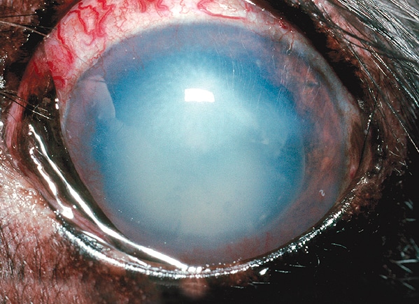
Lens luxation causing conjunctival redness and clouding of the cornea
How is lens luxation diagnosed?
Once the lens completely dislocates it is relatively straightforward to see that it is out of position in most patients. In addition, examination by an ophthalmic specialist using specialised equipment may enable a diagnosis of lens luxation to be made well before the lens actually dislocates out of position.
Is treatment possible?
Lens luxation is a serious, blinding painful condition. Although medical management of primary lens luxation is an option, in most cases surgical removal of the lens is likely to give the best chance of preserving vision. Lens removal may also be indicated if the lens has luxated as the result of other eye conditions, but each of these cases must be assessed on their individual merits.
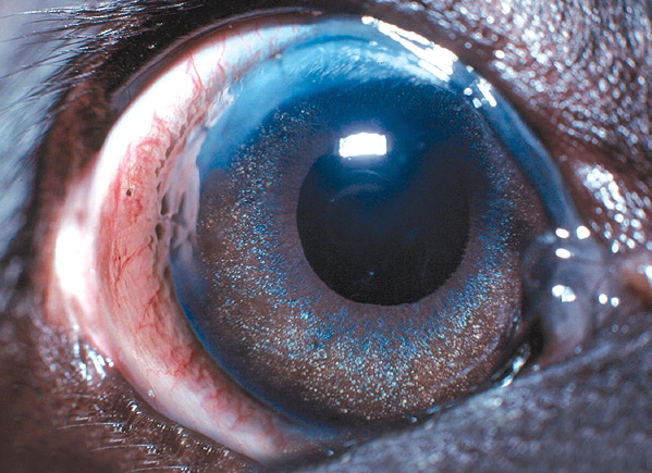
A visual eye following lens removal. Notice the faint scar at the top of the eye
The surgical removal of a luxated lens is an operation that should only be carried out by an eye specialist as it requires special training and skills, the use of an operating microscope and microsurgical instrumentation. The surgery also requires the use of a special muscle-relaxant anaesthetic, as the eye must be held absolutely still in the right position during the operation. During the procedure the patient’s breathing is taken over by the anaesthetist. Again, specially trained staff are required to monitor this anaesthetic.
In surgery for lens luxation, the cornea, which forms the clear window at the front of the eye, is incised and the luxated lens is gently removed. The inside of the eye is coated with special gel during the procedure to help to protect its delicate structures. Some of the natural jelly at the back of the eye (the vitreous) also needs to be removed during the operation, and this is performed using highly specialised mechanised equipment. The wound is stitched with suture material finer than a hair. The stitches dissolve over a period of weeks after the surgery.
Following surgery, intensive medical treatment is required, and several re-examinations are necessary to give the best chance of a successful outcome.
Will my pet cope without a lens?
Most pets cope well without a lens but will need a while to adapt to the new vision. Most dogs without lenses are able to avoid bumping into objects, and many can still chase a ball.
Success chances and possible complications
The chance of a successful outcome depends in part upon the duration of the problem prior to presentation and also whether the lens is completely dislocated at the time of surgery. In early cases, the outcome of surgery can be very satisfactory, but in longstanding cases, the success chances are significantly reduced.
Unfortunately blinding complications are seen in a significant percentage of cases – up to 30% or 40% depending upon the condition of the eye and duration of the problem by the time of surgery. These complications include persistence of glaucoma, bleeding into the eye and retinal detachment.
Even after successful surgery which has saved vision, it may be necessary to continue with long-term medication, especially to keep the pressure in the eye under control. This may in turn require patients to be returned for check-ups from time to time.
Does the condition affect both eyes?
Yes, unfortunately primary lens luxation affects both eyes, although not necessarily at the same time. However, there will often be early signs of partial dislocation of the lens in the second eye at the time that the first eye is noticed to have a problem. Without treatment the second lens will then usually dislocate within a few months of the first, and the natural progression of patients with this condition without surgery is to become blind in both eyes.
Glaucoma then usually develops, and this not infrequently means that one or both eyes may need to be removed in order to relieve pain.
Is preventative treatment available for the second eye?
Yes, especially in cases of primary lens luxation. If the earliest signs of lens instability (subluxation) are detected at the time that the first eye is affected then the lens may be surgically removed. Early removal of unstable lenses carries the best chance of long term success. In early cases it may be possible to perform the surgery through a smaller incision using specialised equipment, and this in turn helps to give a better prognosis.
What happens if vision is irreversibly lost in an eye with a luxated lens?
Eyes that have lost vision irreversibly due to lens luxation are not suitable for lens removal. Because many eyes with lens luxation are painful, it may be necessary to consider removal of the eye under these circumstances.
What happens if an eye needs to be removed?
Eye removal is generally only recommended if the eye is painful and blind and the pain cannot be readily controlled with medication. The thought of an eye being removed may be alarming at first, but it is important to realise that this will generally make the patient feel much more comfortable and much happier very quickly (See Enucleation information page).
The hair is clipped at the site of the operation but soon grows back, and this then leaves hairy skin where the eye used to be. There is no hole left behind, and the result is cosmetically perfectly acceptable. And of course our patients don’t look at themselves in the mirror and worry about their appearance, they just feel more comfortable and get on with life!
If you have any queries or concerns, please do not hesitate to contact us.
Arranging a referral for your pet
If you would like to refer your pet to see one of our Specialists please visit our Arranging a Referral page.