What diseases can affect the inside of the nose?
There are several different types of problem that can affect the nasal cavity of dogs and cats. The most common conditions in dogs include inflammation of the nasal cavity (rhinitis) and nasal tumours (cancer).
Rhinitis in dogs can be due to a number of problems including allergy or unknown cause (called lymphoplasmacytic rhinitis), fungal infection (most commonly a fungus called Aspergillus) and foreign bodies (such as grass blades). In cats fungal disease is rare, whilst polyps (benign outgrowths of tissue) in the nose/throat, rhinitis of unknown cause and tumours are more common. Bacterial infection can occur as a consequence of the underlying disease but usually does not cause nasal disease by itself.
What are the signs of nasal disease?
- Sneezing
- Snorting
- Nasal discharge
- Nose bleeds
- Nasal pain
- Nasal ulceration or loss of pigment around the nostrils
- Reverse sneezing (this can look like spasms of choking or difficulty breathing and can be quite disturbing to see but is not a life-threatening problem)
How is nasal disease diagnosed?
Bloods tests may be performed as part of an initial investigation, looking at general organ health prior to performing an anaesthetic or other tests. Sometimes blood tests are performed to look at how well the patient’s blood is clotting in patients with nose bleeds. Most commonly X-rays or a CT scan of the nasal cavity are performed under general anaesthesia and a procedure called rhinoscopy is carried out, in which an endoscope camera is inserted up the nose or used to look at the back of the throat.
Nasal X-rays may show damage to the small bones in the nose and they may reveal the presence of fluid or tumours. X-rays can sometimes look normal even when nasal disease is present, however.
A CT scan is a much more effective and sensitive way of detecting disease in the nose and may reveal abnormalities that haven’t shown up on an X-ray. A CT scan will also help to define the extent of any damage or the location of a tumour. Rhinoscopy (passing a small camera inside the nose) is performed to look for abnormalities including fungal infection, tumours, and foreign bodies and to help take a tissue biopsy from the nose to send for analysis.
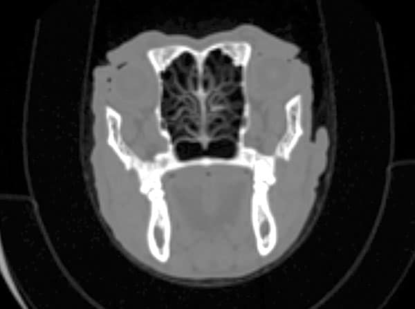
CT scan of a normal nasal cavity, showing a slice across the nose from one side to the other
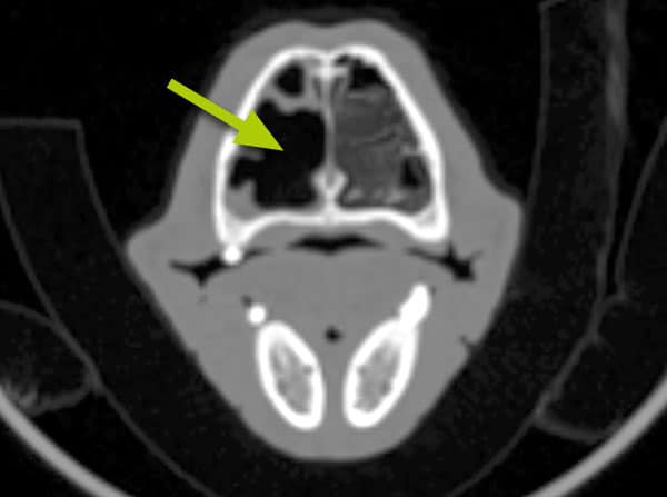
CT scan of fungal infection (Aspergillus): there is destruction of the small bones in the nose, leaving behind an air-filled area which shows as black on the scan (arrow)
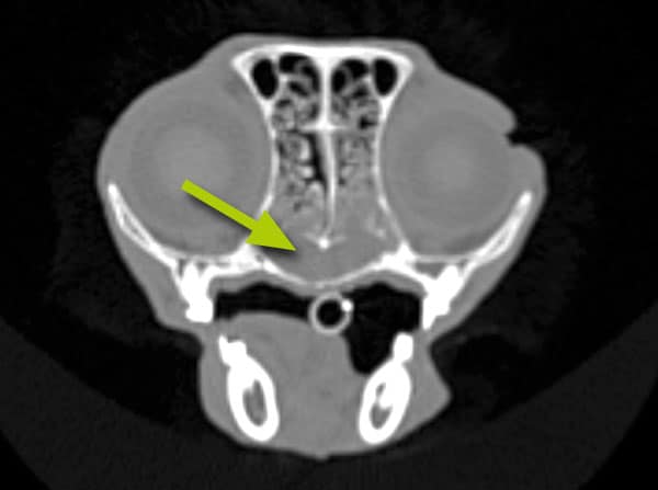
CT scan of lymphoplasmacytic (allergic) rhinitis: there is an increased amount of fluid in the nose. This shows as grey on the scan (arrow)
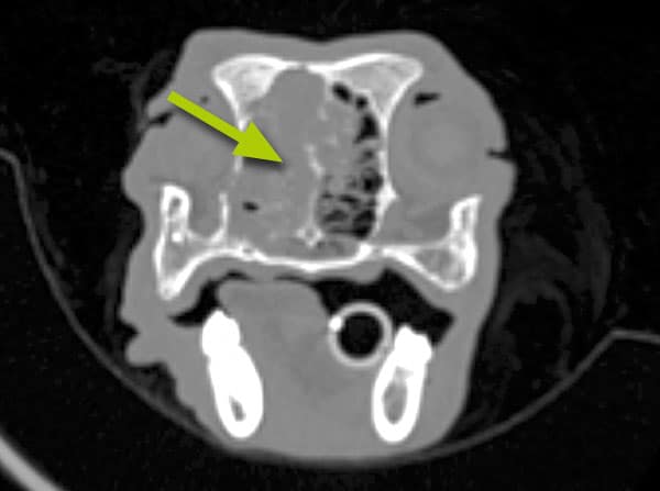
CT scan of a nasal tumour: the tumour (arrowed) has destroyed the bones in the nose)
Rhinoscopy (passing a small camera inside the nose) is performed to look for abnormalities including fungal infection, tumours, and foreign bodies and to help take a tissue biopsy from the nose to send for analysis.
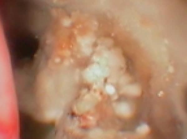
Fungus (Aspergillus) seen as whitish clumps inside a dog’s nose during rhinoscopy (a camera inspection)
How is nasal disease treated?
The treatment for nasal disease depends upon its underlying cause and severity. Sometimes medication in the form of tablets is prescribed; anti-fungal solution must be put into the nose or the sinuses to treat the fungal infection Aspergillus, whilst either radiation therapy or chemotherapy may be required for the treatment of nasal tumours.
What is the long term outlook (prognosis)?
The long term outlook depends upon the underlying disease. Patients usually recover well from removal of a foreign body. Allergic rhinitis is not a life-threatening problem, but treatment is generally aimed at controlling rather than curing the problem and some patients may not respond as well as would be hoped. Aspergillus can respond well to therapy, but the condition may be recurrent and difficult to cure in some patients.
More specific advice and guidance about treatment of nasal disease for individual patients can be given once the diagnosis has been made and detailed information has been obtained from the diagnostic work-up.
If you have any queries or concerns, please do not hesitate to contact us.
Arranging a referral for your pet
If you would like to refer your pet to see one of our Specialists please visit our Arranging a Referral page.