
What is a Total Ear Canal Ablation Surgery?
The term Total Ear Canal Ablation or TECA is used to describe a procedure used to manage severe canal or middle ear disease in dogs where other methods of treatment have failed. The full name of the procedure is Total Ear Canal Ablation with Lateral Bulla Osteotomy (TECA + LBO) but most surgeons use the term TECA for convenience.
The procedure is described in detail below, but a TECA essentially involves the removal of the diseased and infected ear canal whilst leaving the hearing organ itself (the inner ear) in place. The middle ear chamber (tympanic bulla) is carefully inspected by the surgeon at the time of the operation and any abnormal tissue or material is removed. Figure 1 shows the normal anatomy of the outer, middle and inner ear.
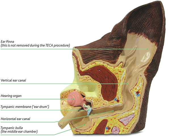
Figure 1 – shows a model of a normal ear canal and internal anatomy
Some useful definitions:
- The ear pinna is the visible part of a dog’s ear. The appearance of the pinna varies between different breeds and can stick up or be floppy in appearance. Most dogs are able to swivel and move their pinna in order to pick up sounds around them.
- The ear canal is an air filled ‘trumpet’ shaped tube that allows sound waves to be channelled from the ear pinna to the ear drum.
- The ear drum is a very thin sheet of tissue that catches the sound waves and transmits them via three very small bones to the hearing organ itself (the inner ear). The ear drum is situated within the tympanic bulla which acts as an amplifying chamber rather like the inside of an acoustic guitar or violin. The tympanic bulla is referred to as the middle ear.
- The hearing organ receives the sound as vibrations and converts it into electrical signals that are then sent to the brain for interpretation. The hearing organ is situated within the skull itself and is referred to as the inner ear.
What causes ear disease in dogs?
Canine ear disease usually occurs due to inflammation of the skin that lines the ear canal (otitis externa) which can lead to secondary infection of the middle ear chamber (otitis media).
In the majority of cases, inflammation of the skin within the ears is part of a more generalised skin condition, with the result that dogs suffering from ear problems often to lick or chew at their feet or experience irritation elsewhere. More severely affected dogs can suffer from clear evidence of skin allergy over other parts of their body, in addition to the ear disease.
The reason that the ears are often more severely affected by this generalised skin irritation than other areas of the body, is because of the environment within the ear canal. Initially the hypersensitivity or skin allergy causes a low level of inflammation which allows bacteria and yeast organisms that normally live on skin to increase in numbers. In mildly affected dogs, most areas of the skin can avoid significant organism overgrowth, but the moist and warm environment within the ear canal provides the ideal environment for these organisms to grow and therefore cause further inflammation. As the organisms increase in numbers, their presence causes further inflammation and damage to the skin, leading to a vicious cycle of deterioration.

Figure 2 – shows a model of an ear suffering from otitis externa (our ear disease) with middle ear involvement (otits media)
As the infection and inflammation progress, the ear canal can become irreversibly narrowed, and the middle ear chamber can also become filled with infected material (Figure 2).
Initial treatment aims to break this vicious cycle using a number of techniques:
- Topical ear antibiotic
- Topical ear anti-inflammatory medication
- Ear cleaner (not always appropriate)
- Systemic (oral) antibiotic and/or anti-inflammatory drugs
- Ear flushing techniques
The key to successful management of ear disease lies in control of the bacteria and yeast organisms within the ear canal and soothing the inflammation, at the same time as addressing the initial cause of the irritation. Treating the initial or underlying cause will usually involve recognition and correction of predisposing factors that may be present in the patient. This may require allergy testing or dietary management to reduce the level of allergic inflammation in the skin.
Ear surgery is reserved for those cases that cannot be managed satisfactorily by medical means.
What happens if I decide to visit North Downs Specialist Referrals for a consultation?
A consultation with a specialist at NDSR can be arranged on a referral basis via your primary care veterinary practice. The initial consultation lasts approximately 45 minutes, during which time you will meet a specialist surgeon from the soft tissue team who will discuss the details of your dog’s medical history and ear disease. During the consultation, potential treatment options for your dog can be discussed in detail.
Unfortunately, often it is not possible to perform a detailed examination of the deeper parts of the ear canal (otoscopic examination/otoscopy) during the consultation, as this generally needs to be carried out under sedation or general anaesthesia, especially when the ears are inflamed and uncomfortable. Final assessment and decisions about treatment therefore usually need to be made following sedation or general anaesthesia, allowing otoscopic examination. Most patients will also require some form of diagnostic imaging e.g. X-rays or a CT scan. If appropriate, the surgeon can then discuss the findings with you over the telephone or in person before proceeding to surgery.
Whilst it is often appropriate to proceed directly from the otoscopic examination to the operating room, it is important to remember that there is never an obligation to proceed with surgery. The surgical specialists at North Downs Specialist Reerrals will always discuss all options with you and an appointment can be made in the future if you require time to consider every option available.
As mentioned above, X-rays or a CT-scan may be required as part of the investigation of ear disease. The requirements and implications of these diagnostic steps, if appropriate, will be discussed at the time of the consultation.
What does the operation involve?
The operation involves the removal of the entire ear canal (Figure 3). The outside part of the ear (the pinna) and the hearing organ (inner ear) itself are left in position. Following removal of the diseased ear canal, part of the bony wall of the tympanic bulla (middle ear) is also removed to facilitate removal of infected material from the middle ear chamber. This step is key to the success of the procedure.
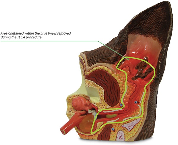
Figure 3 – shows a model of diseased ear and indicates the structures that are removed during TECA surgery
How do I know if my dog will benefit from the TECA procedure?
Your primary care veterinary surgeon will be able to discuss whether your dog may benefit from TECA surgery, and a consultation with a specialist at North Downs can always be arranged through your vet in order to discuss your dog’s ear disease and suitability for surgical treatment.
Chronic ear disease can be very painful for your dog and extremely challenging for both you and your veterinarian to manage. This procedure is designed to eliminate the need for ear drops and significantly improve the comfort level of your dog’s ear. However, as for all surgical procedures, TECA should not be performed without ensuring that it is appropriate. Common reasons for performing TECA include:
- Severe, untreatable or recurrent otitis externa (ear canal inflammation/infection) with minimal respite afforded by medical management
- Ear canal narrowing due to chronic ear disease that has resulted in an inability to adequately medicate the ear canal
- Failed surgery such as Lateral Wall Resection (LWR) or Vertical Canal Ablation (performed to manage ear canal disease)
- Tumours confined to the ear canal
- Otitis externa that has progressed to otitis media (middle ear inflammation/infection) and is non-responsive to medical treatment
- Difficulties in administering topical treatment for ongoing otitis externa
Are there other options?
It is usually appropriate to ensure that all reasonable steps have been taken to manage the condition medially before seeking a surgical solution. Your primary care veterinary surgeon can often help you with this, or a specialist dermatologist (skin specialist) may offer additional conservative management options.
Surgical procedures such as Lateral Wall Resection (LWR) or Vertical Canal Ablation (VCA) are sometimes offered in order to improve the air flow to the remaining ear canal skin and help control the bacterial and yeast populations. These procedures are simpler than TECA surgery and aim to preserve more of the ‘normal’ anatomy of the ear canal. Unfortunately, ear disease inflammation and infection often involves too much of the ear canal for these techniques to be effective and therefore they are not appropriate for many dogs suffering from otitis extern a.
Will my dog still be able to hear after the procedure?
The hearing organ itself is not removed during the TECA operation. However, the removal of the ear canal itself will result reduced hearing sensitivity, similar to the effect of ear plugs or being under water.
Most dogs undergoing TECA have severe changes to the ear canal before the procedure that have already resulted in reduced hearing sensitivity and so most owners do not perceive a large difference post-operatively.
TECA surgery on both sides can sometimes be required and this can result in significant loss of hearing sensitivity. The benefits of the surgery can be profound, but the surgeon will help you weigh this up against the potential reduction in hearing sensitivity.
What is the success rate of this procedure?
When performed by an experienced surgeon, more than 90% of owners report a significant improvement in their dog’s quality of life following the procedure.
What are the potential complications?
Whilst the vast majority of procedures result in success, there are a number of potential complications that can occur. Experienced surgical specialists, such as those at NDSR, will take every step possible to minimise the risk of complications and therefore maximise the chances of a good outcome.
Possible complications include:
Reduced hearing sensitivity – discussed above
Facial nerve paralysis – this can occur in up to 10 to 20% of cases and is usually temporary. The nerve runs very close to the ear canal and can be bruised during the procedure. Paralysis results in some mild drooping of the upper lip and eyelid on the affected side and can result in a reduced blink reflex. Eye lubrication may be required for a time until the third eyelid adapts. Most cases resolve over 4 to 6 weeks.
Bleeding – there are several important blood vessels in the region and significant bleeding can occur during the TECA operation. Experienced surgeons will take steps to avoid significant bleeding and are well positioned to minimise the risk of life threatening levels of haemorrhage.
Wound healing complications – an Elizabethan collar is required to be worn at all times following the procedure until the stitches are removed. The incidence of significant wound healing problems is low, although there is a low risk of infection, as there is with all surgical procedures.
Vestibular syndrome (balance problems) – the organ responsible for balance is positioned next to the organ responsible for hearing. The specialist surgeon will be aware of exactly where these delicate structures are situated and will therefore take every precaution to avoid damage to this region. Unfortunately in some rare cases damage is unavoidable due to the extent of the disease process. Dogs with the complication of balance dysfunction (similar to vertigo) often require significantly more nursing in the immediate postoperative period and some may never fully recover.
Infection/abscess formation- the incidence of this complication is very low when the TECA has been performed by specialist surgeons. Operating in such infected tissue can result in the risk of abscessation which can prove extremely difficult to resolve. Further surgery may be required and not all cases are successfully resolved.
Horner’s syndrome – this is a combination of clinical signs that can result from damage to a complex of nerves that cross the middle ear. Damage to these nerves causes protrusion of the third eyelid, drooping of the upper eyelid and constriction of the pupil of the eye on the operated side. These signs are infrequent in the dog, and are usually temporary. Those cases that do not fully recover do not experience a significant reduction in quality of life or ability to see.
Pinna (ear flap) position and movement – unfortunately the requirements of the procedure can result in an alteration to the appearance or carriage of the pinna in some cases. This may mean that postoperatively the pinna is carried flat rather than upright in some patients.
If you have any queries or concerns, please do not hesitate to contact us.
Arranging a referral for your pet
If you would like to refer your pet to see one of our Specialists please visit our Arranging a Referral page.