What is pemphigus?
Pemphigus is a disease of dogs, cats, horses and goats and belongs to the group of autoimmune skin diseases. These diseases result when the immune system functions abnormally, targeting harmless cells and structures of the body.
In the case of pemphigus, the immune system targets the ‘links’ between the cells of the skin resulting in blisters, pus-filled spots and crusting. There are two main types of pemphigus in domestic animals. Pemphigus vulgaris is a very rare disease and results in deep and painful lesions which can be life threatening. Pemphigus foliaceus (PF) is also rare, but is reportedly the most common autoimmune skin disease in dogs. PF causes more superficial skin lesions and is not normally as severe as pemphigus vulgaris.
PF usually develops in middle-aged to older dogs and cats, although it can commence at any age. In dogs, Akitas and Chow Chows appear to be particularly at risk of this disease, although it can occur in any breed.
What causes pemphigus foliaceus?
PF usually develops due to unknown reasons, although some cases have been linked to drug reactions. It is therefore vital to establish whether any drugs were being used in the months leading up to the development of lesions. Some cases of PF have also been linked to chronic skin disease, as certain dogs with a long history of allergic skin disease have gone on to develop PF. The more serious disease pemphigus vulgaris has also developed due to underlying cancer, so a thorough search for internal disease must be done if this disease is suspected.
What are the clinical signs?
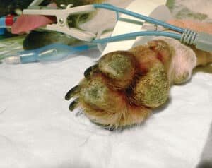
In dogs, PF causes ‘pustular’ lesions. These are pus-filled spots that usually develop into scabs, crusts and sores and are very commonly found on the face, head and ears. Some cases have been described affecting the footpads only (Figure 1), but in more severe cases, the whole body can be affected. Lesions are usually quite symmetrical.
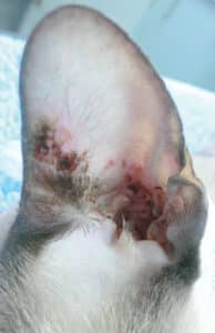
The head, face and ears are commonly involved in PF in cats (Figure 2). Interestingly, cats also commonly suffer from involvement of the claws, and this can sometimes be the most obvious sign. As with dogs, severe cases tend to involve most of the body.
Most cats and dogs remain systemically well, although they can be more lethargic and show a reduced appetite.

Figure 1: Crusting of the footpads in a dog with pemphigus foliaceus
How is it diagnosed?
Based on the animal’s history and clinical signs PF can often be suspected . However, diagnosis can only be made following a biopsy of affected skin. If lesions are present around the face and head, this procedure is usually carried out under general anaesthesia so that the animal remains perfectly still. Only very small biopsies of the skin are needed, and the cosmetic outcome is usually very good.

Figure 2: Crusting around the ear of a young cat with pemphigus foliaceus
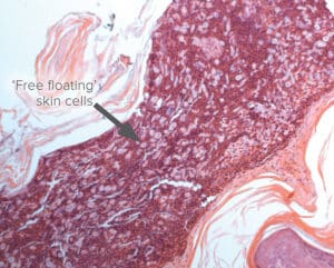
The biopsied skin is sent away for analysis at a laboratory where the pathologist can see if the intercellular ‘links’ have been disrupted within the skin (Figure 3).

Figure 3: Biopsied skin showing the pale free floating cells that have lost their intercellular links in a case of pemphigus foliaceus
How is it treated?
As PF develops due to an ‘overactive’ immune system, treatment aims to reduce the immune attack on the cells of the skin. Steroid medication is the cornerstone of treatment in both dogs and cats and relatively high doses are usually required to control the disease. In most cases, the dose of steroids is then reduced carefully down to a level that controls the disease, but minimises long term side effects. In some cases, other drugs such as azathioprine, chlorambucil and ciclosporin have to used or added in order to achieve control of the disease.
If a drug trigger is suspected, it is also vital to remove this as part of the treatment.
What is the long term prognosis?
PF can usually be controlled quite well, with many dogs and cats remaining free of disease with the use of long term medication.
In the vast of majority of cases, medication will be required in some form for the rest of the animal’s life.
If you have any queries or concerns, please do not hesitate to contact us.
Arranging a referral for your pet
If you would like to refer your pet to see one of our Specialists please visit our Arranging a Referral page.