Syringomyelia is a relatively common condition, especially in breeds like the Cavalier King Charles Spaniel and the Griffon Bruxellois, in which it is suspected to be an inherited disorder. Other names that have been used to describe this condition include syringohydromyelia, Arnold-Chiari or Chiari-like malformation, and caudal occipital malformation.
What is syringomyelia and what causes it?
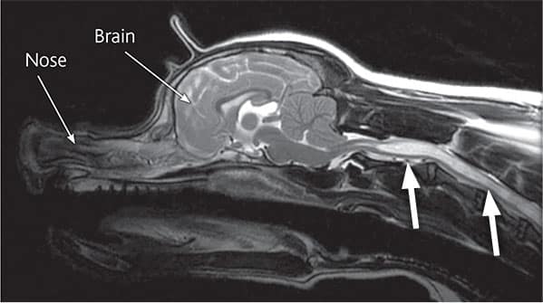
Syringomyelia is a neurological condition where fluid filled cavities develop within the spinal cord (the bundle of nerves that run inside the spine). The most common reason for the fluid build-up is that there is an abnormality where the skull joins onto the vertebrae (the bones of the spine) in the neck, causing fluid in the brain (called cerebrospinal fluid or CSF) to be forced down the centre of the spinal cord, where it causes the tissues to become distended and cavities to form.

An MRI scan of a patient with syringomyelia. The fluid build up in the spinal cord shows as almost white (white arrows).

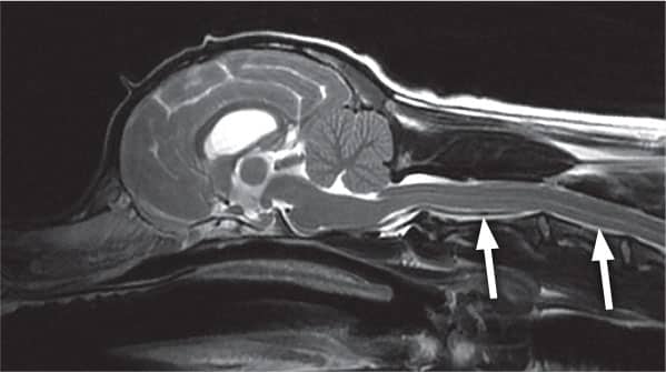
An MRI scan of a patient with a normal spinal cord for comparison (white arrows)
What are the most common signs of syringomyelia?
Clinical signs or symptoms can vary widely between dogs and there is no relation between the size of the syringomyelia (cavity in the spinal cord) and the severity of the signs – in other words a dog with severe fluid build-up can have relatively mild symptoms, and vice versa. The most common symptom that develops is intermittent neck pain, although back pain is also possible. Affected dogs may yelp and are often reluctant to jump and climb. They may feel sensations like ‘pins and needles’ (referred to as hyperaesthesia). Another common sign is scratching of the neck and shoulder region called ‘phantom scratching’, as there is generally no contact of the foot with the skin of the neck. Occasionally dogs become weak or wobbly if there is significant damage to nerves within the spinal cord. Cavalier King Charles Spaniels will typically show clinical signs between 6 months and 3 years of age. Not all dogs with syringomyelia will show signs of pain or other clinical symptoms, so the presence of syringomyelia can be an incidental finding on an MRI scan or specialised X-rays, when neurological investigations are being performed.
Other neurological conditions, such as slipped discs (cervical and thoracolumbar disc disease), can mimic the signs of syringomyelia and it is important for us to rule them out before concluding that your pet is suffering from syringomyelia.
How can syringomyelia be diagnosed?
The best method of diagnosing syringomyelia is an MRI scan of the brain and spine. It is necessary to perform this investigation under a general anaesthetic. The scan and anaesthetic are safe procedures, and patients undergoing an MRI scan for this condition are monitored by our Specialist-led team of anaesthetists. In the future it is possible there will be a genetic test to identify dogs with syringomyelia.
How can syringomyelia be treated?
Medical therapy is usually the treatment of choice in dogs suffering from syringomyelia. Several types of medication are used to manage episodes of pain, including a drug called gabapentin. This drug is safe, with few side effects apart from possible sleepiness. Other medications that may be used include anti-inflammatory drugs, corticosteroids and drugs that reduce the production of fluid in the brain and spinal cord.
Occasionally medical management is unsuccessful and surgery needs to be considered. The aim of surgery is to improve the shape of the back of the skull and reduce the flow of fluid down the centre of the spinal cord. Many dogs will improve following surgery, although some patients will have persistent signs despite surgery, whereas others may show improvement initially but then develop recurrence of their symptoms.
If you have any queries or concerns, please do not hesitate to contact us.
Arranging a referral for your pet
If you would like to refer your pet to see one of our Specialists please visit our Arranging a Referral page.