CT (computed tomography) scanning is a diagnostic tool used to look at various parts of the body, especially those made of bone (including joints), air (including lungs and the nose), and some soft tissue structures, particularly those with a blood supply. CT scanning is used commonly in people, and we use it extensively at North Downs Specialist Referrals. It uses X-rays to produce an image – these are the same X-rays that produce the X-ray images that you may well be familiar with (called radiographs)
CT works by using a continuous beam of X-rays which spins around in a doughnut shaped support called a gantry. The ‘tube head’, which produces the X-ray beam, can spin very quickly, taking as little as half a second to completely circle around the patient. While this is happening, the patient, who is lying on a couch or table, can be moved through the gantry by electronically moving the table top. Each spin of the X-ray tube around the patient in the scanner builds up slices of X-ray images.
After this information is obtained, powerful computers use complex software to produce images that we can recognise and interpret. Interpretation of the hundreds of slices of information, which are obtained for every patient, takes considerable time and expertise.
CT scanning has a number of advantages over both conventional X-ray radiography and some other ways of imaging patients:
- Because it produces slices which are cross-sections of the patient, CT scanning removes superimposition by overlying structures, making interpretation of complex parts of the body a lot easier, hence allowing us to make more difficult diagnoses. By contrast, X-ray radiography effectively takes an image as a semi-transparent ‘shadow’, and structures are seen superimposed on top of each other, making it more difficult to interpret any abnormalities.
- An advantage that CT has over all other imaging modalities is that the slices can be added together electronically to give much thicker slices. In this way, we often see small abnormalities that might otherwise have been missed (by improving something called ‘signal to noise ratio’ for those who are interested!).
- As opposed to MRI scanning, the slices from a CT scan can be electronically stacked up in any direction, allowing the radiologist (the imaging Specialist vet) to manipulate the images and effectively, reconstruct, the tissues. This can often help surgeons to understand disease better, as they can have a more three dimensional ‘surgeon’s eye view’ of the abnormalities.
What is CT used for at North Downs Specialist Referrals?
At NDSR, CT scanning is most often used for examining noses, lungs, the contents of the abdomen, and bones. CT has revolutionised the way the veterinary profession looks at problems within complicated joints such as the elbow, for example.
Here are some examples of how useful CT can be:
Joints
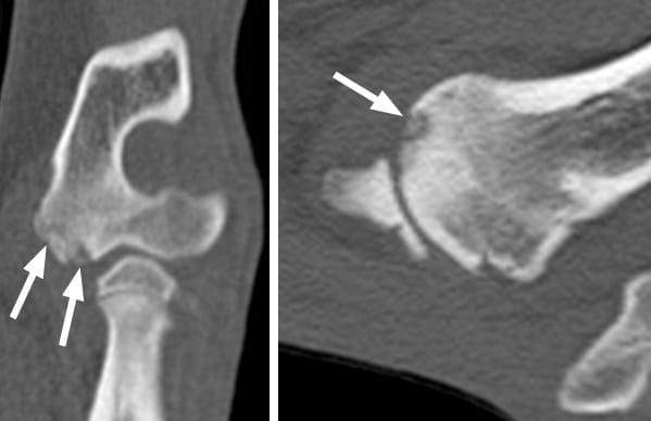
CT scanning is particularly useful at looking at complex joints (those that are difficult to fully assess with normal X-rays, or are composed of more than two bones). CT is used extensively to evaluate the elbow joint, as it is notoriously difficult to assess by radiography.

On these CT scans you can see a small erosion (arrows) on the end of the humerus (the bone of the upper limb) just above the elbow joint. This is a disease called elbow osteochondrosis, often seen in young animals. This would have been very difficult or impossible to see on normal X-rays.
Nose
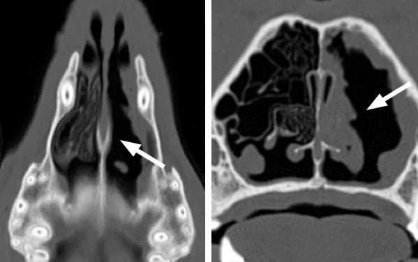
CT scanning has largely replaced normal X-rays for looking at diseases of the nose. This is because normal X-rays are not very sensitive at looking at nasal disease (that means they often miss the disease that is present) and even worse, can often suggest disease is present when it is not. Nasal cancer is often assessed by CT, in addition to a host of other diseases such as fungal infections, cysts etc.

These CT scan images show two views (from above and end-on) of a disease called destructive rhinitis, most commonly caused by a fungus that invades the nose. You will see that the nasal cavity on the left of each image looks normal, with lots of normal scrolled structures inside the nose called turbinates. On the other side, these delicate structures are almost entirely absent (arrows), having been destroyed by the fungus.
Chest
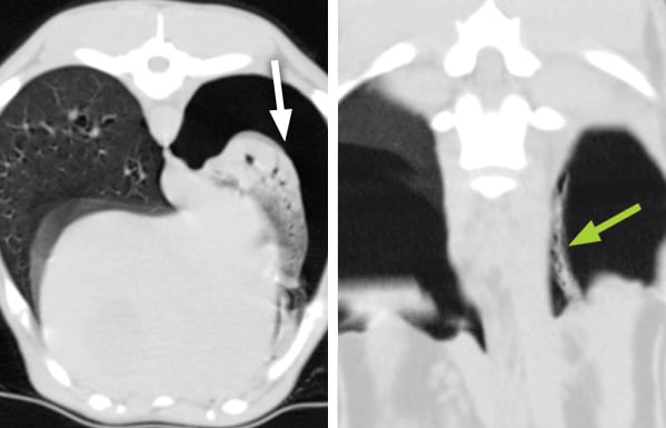
CT scanning is extremely useful at looking at the chest, particularly for those structures filled with air (the lungs and the windpipe or trachea). It has revolutionised the way we detect diseases that may have been very difficult to assess on normal X-rays.

The image on the left shows a relatively normal lung (on the left of the image) and a lung that is collapsed (white arrow). The collapsed lung is also affected by pneumonia. In the image on the right, a small straight lesion was found loose in the cavity in which the lung is housed. This was a grass seed (green arrow) which was removed surgically by one of NDSR’s soft tissue surgical Specialists. This grass seed would not have been seen on a normal X-ray.
Abdomen (tummy)
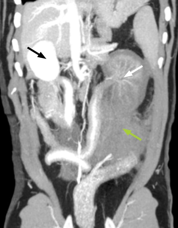
CT scanning is very useful for looking at soft tissues within the abdomen because it is quick (much faster than MRI) and is able to spot changes that can be seen after contrast (dye) is injected (see above). Using contrast agent is very useful for looking at abnormal blood vessels in the abdomen, such as those seen in a condition called a portosystemic (liver) shunt (see our Portosystemic Shunts Information Sheet), as well as for many other diseases.

This CT image of a dog’s abdomen, obtained after injecting contrast agent (dye), shows a normal right kidney (the white structure on the left of the image – black arrow) and a kidney on the right of the image that has a reduced blood supply (white arrow). This dog had been hit by a car with the result that the left kidney has been torn off its blood supply – the normal contrast or dye (which shows as white) is not present in the abnormal left kidney to the same degree as in the normal right kidney. The grey material (green arrow) near the affected left kidney is blood from the severed artery. The patient made a full recovery after surgery.
If you have any queries or concerns, please do not hesitate to contact us.
Arranging a referral for your pet
If you would like to refer your pet to see one of our Specialists please visit our Arranging a Referral page.