
MRI (magnetic resonance imaging) scanning is a diagnostic test that is used to look at various parts of the body, including the brain, spine, eye sockets, joints and soft tissues. MRI scans are commonly used in people, and they are also frequently performed at North Downs Specialist Referrals. Unlike X-rays, MRI scanning uses a magnetic field rather than radiation.
During an MRI scan the patient is placed in a magnetic field, and this temporarily alters the atoms of hydrogen in the body. As the magnetic field is altered, parts of the hydrogen atoms change their alignment, giving off faint radio waves which are then picked up by sensitive, specially designed coils placed around the part of the patient that is being imaged. A powerful computer then analyses the signals that are received by the coils, and this builds up a picture of the parts of the body which are being investigated. Sophisticated changes can be made to the magnetic field and radio frequencies to highlight different tissues and different diseases within the tissues.
An MRI scan is made up of lots of images which are obtained as slices or cross-sections of the patient. These slices allow individual structures to be imaged without having to look at them through other structures that might get in the way (this superimposition of structures is a drawback of traditional X-rays or radiographs). The direction of the slices can be changed to give the best information about any particular structure.
Brain
As the brain is surrounded by a hard bony case (the skull), other types of imaging such as X-rays struggle to produce useful images of the brain tissue. Computed tomography (CT scanning) can be used to image the brain, but the images are not as clear as with MRI. In very young patients, or animals with skull defects such as open fontanelles (the ‘soft spot’ on the skull in newborn infants), an ultrasound scan can be used to image the brain, but the view obtained is limited and can be difficult to interpret.
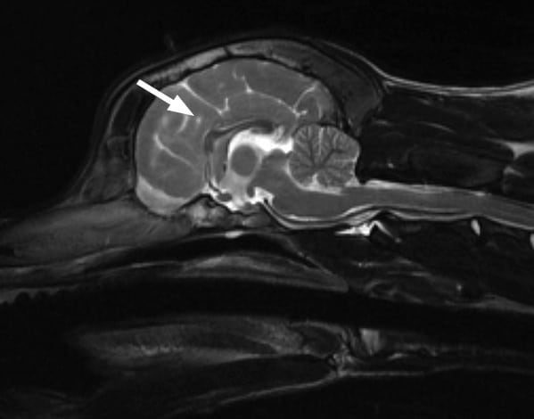
An MRI scan of the brain (arrow), showing the detail which can be seen – in this scan the cross section of the brain seen is viewed from the side
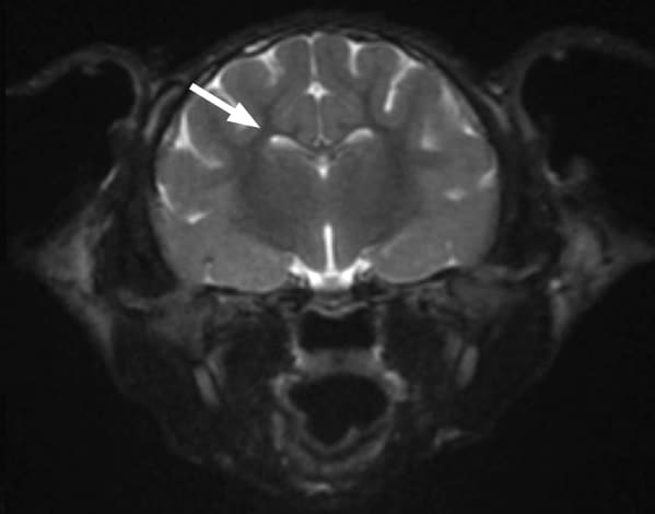
Another MRI scan of the brain (arrow) This view is a cross-section seen from the back
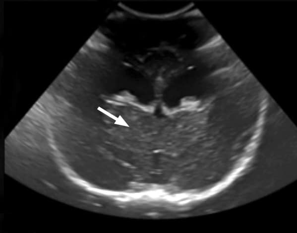
An ultrasound scan of the brain (arrow) through the soft spot on a skull – the detail seen is very limited compared to the MRI scan. This view is a cross-section seen from the back
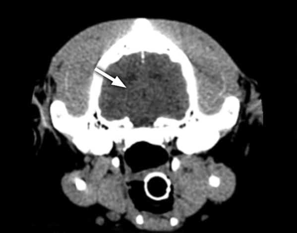
A CT scan of a brain (arrow), again showing much less detail than the MRI scan. This view is a cross-section seen from the back (the skull around the brain is shown as bright white)
Spinal cord
Traditionally conventional radiography (X-ray) has been used to look at the spinal cord.
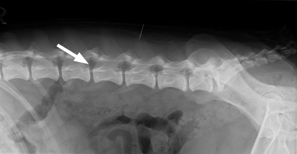
A normal X-ray of the spine – the bones are clearly visible, but the area of the spinal cord (arrow) shows no soft tissue detail
CT scanning shows up the bones of the spine very well, but provides only limited information about the nerves of the spinal cord itself. The same dye that is used in an X-ray myelogram can also be injected around the spinal cord when a CT scan is performed. This provides much more information compared with conventional X-ray myelography, but the risks associated with injecting contrast agent around the spinal cord remain.
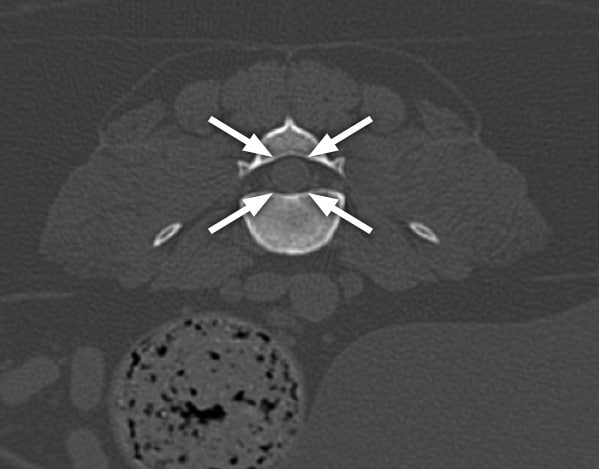
A CT scan of the spine showing a cross section through a vertebra (seen as white) with the spinal cord (arrowed) in the middle of the bone
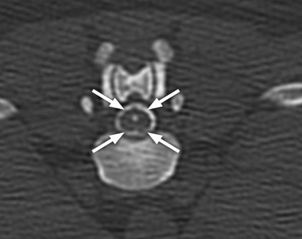
A CT myelogram with dye (seen as white) injected around the spinal cord (arrows)
In comparison, MRI provides superb detail of the spinal cord and surrounding soft tissues in a safe, non-invasive fashion.
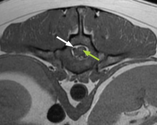
MRI image of the spine showing abnormal material (a ‘slipped disc’ – white arrow) next to the spinal cord (green arrow), with no need for dye injection
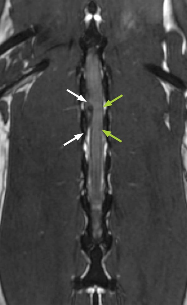
MRI scan of the spinal cord (this time in a slice viewed from above), showing slipped disc material (white arrows) bulging out and pressing against the spinal cord (green arrows), with no need for dye injection
If you have any queries or concerns, please do not hesitate to contact us.
Arranging a referral for your pet
If you would like to refer your pet to see one of our Specialists please visit our Arranging a Referral page.