Ultrasound scanning is a diagnostic test that is used to look at various parts of the body, in particular the heart, the abdomen (tummy) and other soft tissues (as opposed to bones). Ultrasound is commonly used in people (most familiarly in pregnancy), and is performed at North Downs Specialist Referrals on a daily basis. Unlike X-rays, ultrasound scanning uses sound waves rather than radiation to obtain an image.
During an ultrasound scan, the patient is placed on a table and gently held. An ultrasound probe, which looks a bit like a microphone, is held against the part of the body being imaged. The probe transmits and receives ultrasound waves, and a powerful computer then analyses the waves and builds up a picture of the parts of the body which are being investigated.
Ultrasound waves do not pass through air and, because of this, the patient must be prepared carefully for the examination. The patient’s hair must be clipped in the region that is being examined, and a gel is applied to both the skin and the probe, to ensure good contact between them.
Ultrasound scanning is a diagnostic imaging technique which provides superb detail in soft tissues (as opposed to bones). An ultrasound scan allows examination of parts of the body which are not easily seen on X-rays, such as the internal structure of the heart and eye, the contents of the abdomen (tummy) and the muscles in the limbs. Normal X-rays (radiographs) cannot distinguish between the outside wall and the inside cavity of hollow, fluid-filled organs, such as the bladder, whereas ultrasound allows these two separate parts to be identified and examined in detail. At NDSR, ultrasound scanning is used on a daily basis, with over 1000 such examinations performed each year.
Ultrasound scanning provides a moving ‘real time’ image, a bit like an instant movie of what the probe is ‘seeing’. This allows assessment of movement in the heart or other structures in the body, and it also enables the imaging Specialist to ‘piece together’ the images in their mind as the probe is moved around. Movement of blood in the heart and blood vessels, as well as in organs, can also be assessed. Real time imaging also allows the experienced imaging Specialist to take samples (biopsies) of different tissues under ultrasound guidance, where the sampling needle can be directly visualised with ultrasound in the tissues at the time the sample is being taken.
Is there anything special about the CT scanner and imaging personnel at North Downs Specialist Referrals?
The short answer is, yes! Not only does NDSR have sophisticated ultrasound scanning equipment, but the imaging department is staffed by a highly experienced team of accredited, recognised Specialists.
Further examples of the use of ultrasound scanning at NDSR
Abdomen
The majority of ultrasound scans performed at NDSR are of the abdomen (the tummy). Ultrasound provides superb detail about the internal structure of the organs, without the need for any dyes to be injected into the patient, unlike X-ray or CT scan contrast studies. Ultrasound allows differentiation of fluid and solid tissue and is invaluable when there is abnormal fluid (called ‘ascites’) within the abdomen. In addition, ultrasound allows assessment of movement in real time, so stomach and gut movement can be seen, as can urine entering the bladder from the kidneys. Sampling of the different tissues and fluids within the abdomen can be performed with increased safety under ultrasound guidance, rather than being carried out ‘blind’.
A video ultrasound scan of urine entering the bladder. The urine enters the bladder via the tubes from the kidneys (the ureters) and it can be seen squirting into the bladder as the red coloured jet (on this Doppler colour-flow scan the red colour indicates a rapid flow of fluid)
Chest
Although ultrasound waves do not pass through air (and therefore not through normal air-filled lungs), ultrasound scanning can be used to examine abnormal chests when a disease resulting in fluid build-up or a mass (lump) is present. As with the abdomen and other regions, any abnormal areas seen on the scan can be sampled using ultrasound guidance, where appropriate. In addition, ultrasound is the diagnostic imaging technique of choice for examination of the heart, allowing the heart chambers and valves to be seen in great detail, as well as enabling blood flow through the heart chambers to be assessed.
A video ultrasound scan of the heart beating – the image shows a cross section of the left ventricle at the level of the muscles that attach to the heart valves.
Eye
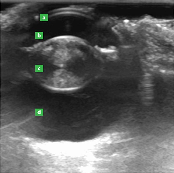
As the eye contains mostly jelly and no bones or air, ultrasound is ideal for demonstrating changes within the eyeball itself. Again, samples can be taken from the eye and surrounding structures under ultrasound guidance.

An ultrasound image of an eye showing a cataract in the lens (c). The cornea (a) and the fluid/jelly-filled segments of the eye (b and d) are also visible.
Muscles
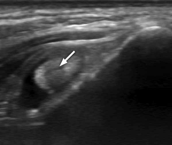
Ultrasound is also used to assess the soft tissues of the limbs, such as muscles or ligaments. Torn or injured soft tissues, for example, tendons, can be assessed with ultrasound and healing can also be followed by repeating the scan at intervals.

Ultrasound image of an abnormal biceps tendon (arrow) with fluid around it (showing as black).
If you have any queries or concerns, please do not hesitate to contact us.
Arranging a referral for your pet
If you would like to refer your pet to see one of our Specialists please visit our Arranging a Referral page.