
What is the atlanto-axial joint?
The atlanto-axial joint is the joint between the first and second cervical bones (vertebrae) in the neck, referred to as the atlas and axis respectively. It differs from the joints between the other vertebrae in that there is no ‘disc’ present. Instead, the atlanto-axial joint is stabilised by ligaments just like those in the joints in a leg, for example. The design of this joint allows the dog’s head to move from side to side.
What is meant by atlanto-axial subluxation?
Subluxation of the atlanto-axial joint occurs when the normal alignment of the first and second vertebrae in the neck is disrupted. This results in excessive movement of this joint (instability) which can cause neck pain and pressure on the nerves of the spinal cord which runs through the tunnel (vertebral canal) created by the vertebrae. Although an injury can result in damage to the ligaments or the vertebrae, it is more common that dogs are born with an abnormality of the second cervical vertebra (the axis) or of the ligaments, which makes the likelihood of subluxation developing greater, often as a consequence of minimal, (if any,) trauma.
Could my dog have atlanto-axial subluxation?
Although any dog could develop atlanto-axial subluxation due to trauma (for example, running into a patio door), it is most commonly seen in toy breeds of dogs, such as Yorkshire Terriers or Poodles. Very rarely it can occur in cats. Often in toy breeds of dogs, a congenital abnormality underlies the problem, and for this reason the condition is most often seen in immature dogs (less than 1 year of age). Affected dogs may show signs of neck pain, such as yelping and crying or rigidity of the neck. More subtle signs may include a low head carriage or difficulty lowering the head to eat or drink. Signs of spinal cord injury can vary in severity, from mild weakness or incoordination in all four limbs to inability to stand and walk.
How is atlanto-axial subluxation diagnosed?
A detailed neurological examination is necessary to detect evidence of spinal cord injury and possible neck pain. Investigations are then required to confirm atlanto-axial subluxation and distinguish it from many other neck problems. Investigations usually require a general anaesthetic and this must be undertaken with extreme care, as manipulation of the neck can exacerbate any spinal cord injury.
Normal X-rays (radiographs) of the neck can be very useful for diagnosing atlanto-axial subluxation, but they provide only limited information about any underlying bone deformity and no information regarding the severity of any spinal cord injury. Sometimes more advanced imaging techniques such as an MRI or CT scan are required. A CT scan (a 3-dimensional X-ray) can confirm the diagnosis and provide more detailed information about any bone deformity, which in turn can help with the detailed planning required for a potential surgical procedure. MRI scanning uses high powered magnets and a computer to generate images of the spine. MRI can also confirm the diagnosis, help in assessing any spinal cord injury, and it can rule out other brain or spinal cord abnormalities that may be present. In some situations a combination of X-rays, MRI and /or CT may be required.
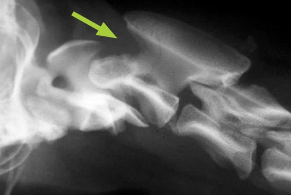
X-ray of the neck in a miniature breed dog showing separation of the first two bones in the neck (atlanto-axial subluxation or instability), leading to an increased space between them (arrowed)
In some patients it may be necessary to collect some fluid from around the spinal cord (cerebrospinal fluid – CSF) and send it to a laboratory for analysis. This test assists in ruling out a diagnosis of an inflammatory condition of the spine – these types of disorder are also seen most commonly in immature dogs. If this test is required, a ‘lumbar puncture’ at the base of the spine (as is carried out in humans) is performed, as this avoids excessive manipulation of the neck.
How can atlanto-axial subluxation be treated?
The two ways to manage atlanto-axial subluxation are with (1) conservative treatment or (2) surgery.
- Conservative treatment
This involves strict cage rest, application of a neck brace and giving painkillers. Maintaining a neck brace, often for several weeks, is very difficult and is poorly tolerated by many patients. - Surgery
The aim of surgery is to stabilise (fuse) the atlanto-axial joint in a normal position. This alleviates neck pain and enables the spinal cord to recover from injury. A number of surgical techniques can be used, but the most reliable approach involves placing bone screws into the first and second cervical vertebrae (the atlas and axis) and connecting the screws together with special bone cement. The surgery is performed through an incision made on the underside of the neck. It is a very intricate procedure due to the location of the problem and the small size of many of these patients. Atlanto-axial subluxation surgery should only be performed by experienced surgeons with advanced training.
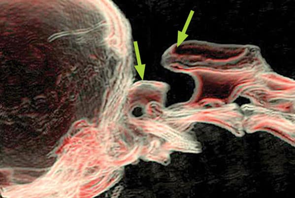
CT scan showing two vertebrae in the neck (the atlas and axis) partially separated before surgery (arrows)
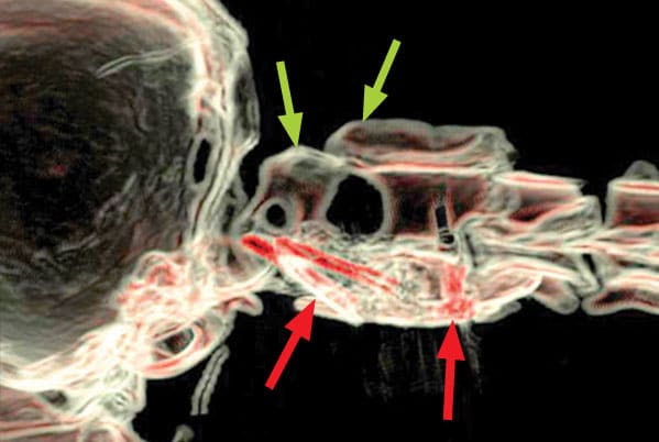
CT scan after the operation showing the two vertebrae back together again (blue arrows) The screws are visible and show as red on this image (indicated by red arrows) and they are supported by a plug of cement under the bones
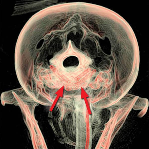
A CT scan looking from behind the skull, showing the screws in the bones from a different angle (indicated by arrows)
What is the outlook with atlanto-axial subluxation?
The outlook (prognosis) with atlanto-axial subluxation depends on how it is managed.
With conservative treatment, although an improvement can be expected in many cases, pain and spinal cord injury often recur once the neck brace is removed and activity is increased. As a consequence, surgical stabilisation of the atlanto-axial joint is indicated in most patients.
The success rate with surgery is generally good provided the atlanto-axial joint is adequately realigned and screws and cement are placed in the bones in a safe manner. The prognosis tends to be better in dogs that show clinical signs whilst still young, can still walk prior to surgery and where signs have not been present for a long time.
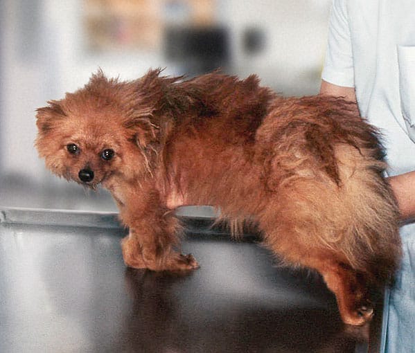
A young Chihuahua before surgery for atlanto-axial subluxation
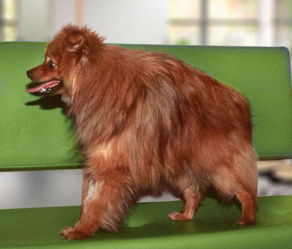
A young Chihuahua after surgery for atlanto-axial subluxation. Stabilising the bones at the top of the neck abolished pain and enabled this small dog to walk again
Arranging a referral for your pet
If you would like to refer your pet to see one of our Specialists please visit our Arranging a Referral page.