
What is osteosarcoma?
Osteosarcoma is a cancer that originates in bone tissue. In dogs the most commonly affected bones are the humerus (upper arm), radius/ulna (forearm), femur (thigh) and tibia (shin). Other bones, such as those of the skull, spine, scapula (shoulder blade) and pelvis, are less frequently affected.
Large and giant breeds are much more likely to develop an osteosarcoma than medium and small breeds. In dogs, osteosarcoma affecting the long bones tends to be highly malignant with spread (metastasis) of cancer cells to other parts of the body especially the lungs. This has typically occurred at a microscopic level in most dogs by the time the tumour is detectable.
What are the signs of osteosarcoma?
Osteosarcoma affecting the humerus, radius/ulna, femur or tibia will typically cause severe bone pain and associated fore or hind limb lameness. The lameness tends to be progressive, relentless and responds poorly to conventional pain killing drugs. As the tumour grows it may result in a visible or palpable swelling of the affected bone. Muscle wastage (atrophy) is often a marked feature. In advanced cases the cancer may weaken the bone to such an extent that it fractures.
How is osteosarcoma diagnosed?
Radiography (X-ray) is the most common method of diagnosing osteosarcoma. Evidence of bone destruction is a common feature, and when detected it is highly suggestive of osteosarcoma, especially since other bone tumours and other conditions that cause bone destruction, such as bone cysts and bone infections, are much less common. Advanced imaging techniques, for example a CT scan, may provide additional information, including the extent of bone involvement. Radiographs or a CT scan of the chest and an ultrasound scan of the abdomen should be considered in dogs with suspected osteosarcoma to look for evidence of metastases (tumour spread). A definitive diagnosis of osteosarcoma requires a sample of tissue being obtained (a biopsy) and examined microscopically. Blood tests may also be performed to assess the patient’s general health and fitness for treatment.
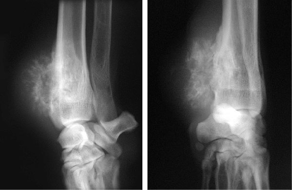
X-rays showing both destruction of bone and irregular new bone formation in a Great Dane with an osteosarcoma affecting the end of the radius (bone in the forearm)
What are the treatment options for osteosarcoma?
The main treatment option for osteosarcoma affecting the long bones in dogs is the combination of surgery and chemotherapy. Amputation of the affected limb provides rapid pain relief and is a good option in those dogs that have no significant problems with their other three limbs. Even giant breed dogs rapidly adapt to losing a limb, and client satisfaction about their dog’s wellbeing is generally high following this type of surgery. The possibility of local recurrence of tumour is minimal with this approach. However, spread to other parts of the body is very common, with the result that very few dogs undergoing amputation alone remain alive six or more months after surgery. In order to achieve a more appreciable life expectancy, surgical removal of the tumour is followed by chemotherapy. Using the combination of amputation and chemotherapy, almost 50% of patients will still be alive one year after diagnosis.
Surgical alternatives to amputation include techniques which remove the tumour but maintain the limb (referred to as ‘limb sparing’). The cancerous section of bone is removed, and metal plates and screws are used to bridge the gap which is filled with a graft or prosthesis. Fusion (arthrodesis) of the adjacent joint is usually required, because osteosarcomas tend to involve the extreme ends of bones near joints. Limb function following removal of a tumour affecting the end of the radius accompanied by fusion of the carpal joint (wrist) is generally very good, with average survival times similar to those following amputation. However, there are significant risks associated with limb-sparing surgery, including the risk of cancer recurrence, implant breakage and severe, persistent infection. Chemotherapy is just as important in the management of these limb-sparing surgery candidates as it is in amputees. It is very important to remember that the invisibly small cancer spread will still progress if no measures are taken to control it.
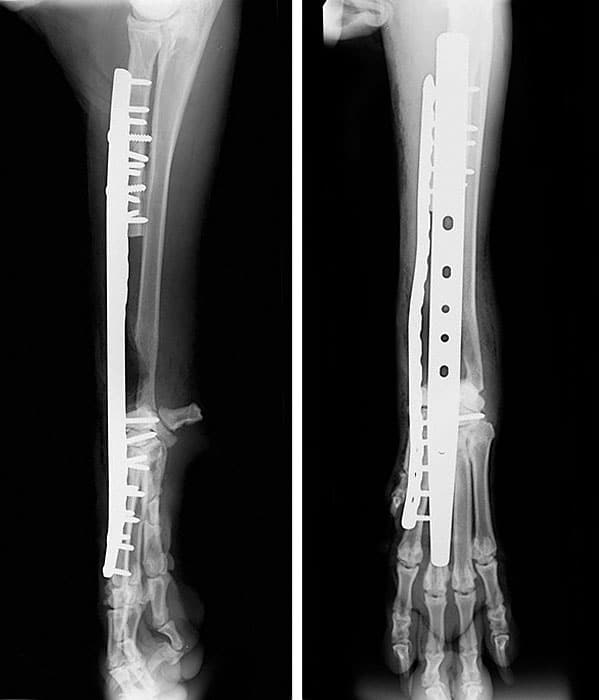
X-rays obtained after surgery in a Newfoundland with an osteosarcoma affecting the radius. The tumour has been excised (cut out) and the gap in the bone bridged with two plates that are attached to the remaining bones with multiple screws. The carpus (wrist joint) has been fused.
Clinical Trials:
There is a steadily growing body of evidence that the immune system can play a significant role in the management of the microscopic metastases (spread of cancer) in patients with osteosarcoma. We currently have a clinical trial available which investigates the role of an immunotherapy product, which is implanted directly into the tumour prior to tumour removal. Eligible patients must have a suspected or proven osteosarcoma of a limb bone. They must be candidates to undergo amputation or limb-sparing surgery and chemotherapy. Apart from the immunotherapy product, patients would receive the same care as any other patient with osteosarcoma. No patients would be randomised to receive a placebo (a non-active medicine substitute). Upon completion of the study, we hope the results will inform the development of a much wider, international trial of the same product.
Non-Surgical Treatments:
Some patients are considered not to be candidates for surgery. Pain-killing treatment is important in all of these patients. In addition, radiotherapy might be recommended, with or without chemotherapy. Radiotherapy does result in an improvement in comfort in the limb in approximately 70% of cases. This effect is variable in duration but typically lasts two to four months. The duration of effect is improved with additional chemotherapy.
Over the years, there has also been a reasonable amount of work done looking at drugs which are supposed to increase the density of bone, thus making it stronger. These drugs are called bisphosphonates. The evidence supporting their use is extremely weak and there are now good studies that show no effect whatsoever. However, there is a chance that these treatments might be beneficial in individual patients. We are always happy to discuss all options and will administer treatments with a low likelihood of success if owners truly understand the slim chance of benefit and any potential risks that might be involved.
Osteosarcoma at other sites:
When osteosarcoma affects the mandible (lower jaw bone), maxilla (upper jaw bone), skull or nasal cavity (nose) the treatment typically comprises surgery, with or without radiation therapy. Osteosarcoma affecting these sites may have a better long-term outcome, but this varies widely depending on the site and size of the tumour.
If you have any queries or concerns, please do not hesitate to contact us.
Arranging a referral for your pet
If you would like to refer your pet to see one of our Specialists please visit our Arranging a Referral page.