
Canine appendicular limb osteosarcoma occurs predominantly at four sites which, in order of prevalence are: distal radius, proximal humerus, distal femur and proximal tibia.
A massive 22% of diagnoses are reported to arise in the proximal humeral metaphysis (versus 29% in the distal radius). Furthermore, it is our observation that the proximal humeral location accounts for an increasing proportion of the overall cohort. While this may simply reflect changes in referral behaviour, it is interesting to speculate that it might reflect a genuine epidemiological change, perhaps due to a change in dog ownership habits. Certainly, greyhound ownership has reduced considerably with the decline in greyhound racing in recent times.
The typical presenting history for a dog that proves to have osteosarcoma comprises an unseen injury that occurred at exercise, followed by almost complete recovery after a period of rest and anti-inflammatory medication and further progression once normal exercise resumes. The apparent relapse does not respond to rest and medication.
In cases of forelimb lameness due to distal radial osteosarcoma, once cancer is suspected, it is quickly recognised due to subtle asymmetry of the craniomedial antebrachia at this time. Therefore, given its predilection for specific metaphyseal sites, if osteosarcoma is suspected but there is no indication of a distal radial problem, a very simple diagnostic test is to press your thumbs firmly into the proximal humeral metaphysis from a craniolateral approach. This can elicit an extreme response so please proceed cautiously.
Radiography is indicated in dogs with a chronic lameness that cannot be localised to a specific region of the forelimb. It would be reasonable to radiograph the whole limb. It is good practice to radiograph both limbs so that a template image is available to help interpretation of features that might or might not be significant.
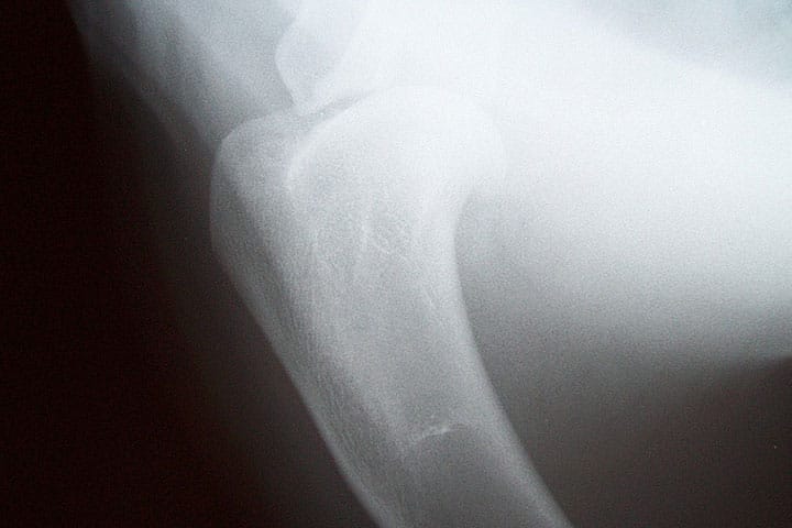
The classical radiographic appearance of proximal humeral osteosarcoma is characterised by the combination of cortical bone loss, evidence of periosteal proliferation, and an indistinct margin between normal and abnormal bone. Sometimes these lesions can be very subtle. Superimposition of other thoracic structures can limit your ability to interpret the area of greatest concern. Localisation of osteosarcoma to the proximal humeral metaphysis is incredibly consistent, so if the area of interest cannot be evaluated confidently, consider repeating the radiographs with the forelimbs more protracted or consider sending the radiographs to us for an additional point of view. In some cases we just cannot be sufficiently confident using radiography and in these circumstances, CT would be recommended.
Case Advice or Arranging a Referral
If you are a veterinary professional and would like to discuss a case with one of our team, or require pre-referral advice about a patient, please call 01883 741449. Alternatively, to refer a case, please use the online referral form
About The Discipline
Oncology

Need case advice or have any questions?
If you have any questions or would like advice on a case please call our dedicated vet line on 01883 741449 and ask to speak to one of our Oncology team.
Advice is freely available, even if the case cannot be referred.
Oncology Team
Our Oncology Team offer a caring, multi-disciplinary approach to all medical and surgical conditions.