Patients with respiratory disease commonly require anaesthesia in practice. These patients can be challenging to anaesthetise and recover safely. The following Q&A answers some of the common questions and concerns regarding these patients.
What are the most common reasons for a cat or dog in respiratory distress to require anaesthesia?
- Trauma
- Obstruction of the airways
- Diaphragmatic hernia (congenital or traumatic)
- Lung lobe torsion
- Pneumonia
- Bronchial disease
- Asthma
- Lung oedema
What are the abnormal respiratory patterns we can detect during physical examination?
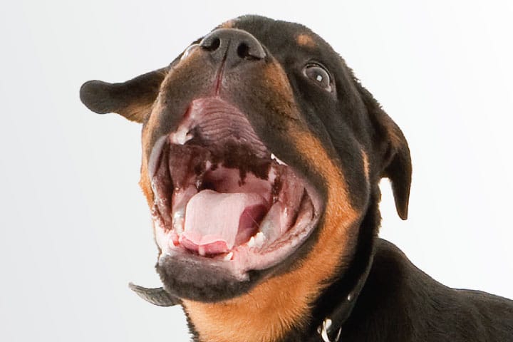
Animals with respiratory disease might present an abnormal breathing pattern characterised by polypnea or dyspnoea with or without abdominal effort. Dyspnoea can be further characterised in inspiratory, expiratory or both.
Orthopnea is present in animals that struggle to ventilate when recumbent. When animals suffer from raised intracranial pressure, they can present with abnormal breathing patterns such as Cheyne-Stokes breathing: periods of apnoea followed by periods of increased ventilation. Paradoxical abdominal movement (chest and abdomen move in dys-synchronous way) is seen with pleural space disease. Respiratory sounds include inspiratory stertor (upper respiratory tract obstruction), inspiratory stridor (laryngeal and extrathoracic tracheal obstructions), expiratory stridor (intrathoracic tracheal collapse).
Sometimes animals present with no respiratory signs at all.
How important is imaging before anaesthesia?
Obtaining a view of the chest before anaesthesia will help narrow down the list of differential diagnoses and allow an estimation of the degree of lung tissue compromise. Although a radiographic view of the chest is often recommended, a relatively new ultrasound technique of the chest provides rapid, stress-free access to the pleura, the superficial lung parenchyma and the heart. These techniques are called T-FAST and VetBlue and you can learn more about them at https://fastvet.com.
What are the considerations before choosing a premedication?
- Severity of clinical signs
- Differential diagnoses
- Age, behaviour, co-morbidities of the animal
- Familiarity with drugs
- Availability of drugs
In general, we recommend avoiding major sedatives and opt for an opioid-only premedication, unless the animal is unco-operative.
What are the main considerations during induction?
During induction the animal might become apnoeic or decrease there tidal volume and/or respiratory rate. As the body has no oxygen reserves (and even more so if it starts with an oxygen debt), this can lead to rapid desaturation. Preoxygenation in this case is fundamental to avoid desaturation. This is achieved with a tight fitted mask for 4-5 minutes before induction. If the animal does not tolerate this, they can be placed in an oxygen-filled incubator before general anaesthesia (size and temperature allowing) or they can receive oxygen via flow-by, although this method will likely not achieve a high percentages of inspired oxygen.
The induction process must be rapid to allow rapid control of the airways, delivery of oxygen and manual or mechanical ventilation.
What should we focus on monitoring during anaesthesia of dogs and cats with respiratory disease?
- Respiratory rate and pattern
- Saturation of haemoglobin with oxygen (SpO2)
- End tidal carbon dioxide (etCO2)
More complicated cases will require:
- Spirometry (to assess lung volumes and pressures changes during anaesthesia)
- Blood gas monitoring
I have been told to use conservative fluid therapy in animals with respiratory disease, is this true? Why?
It is recommended to use low fluid rates in people with lung oedema and ARDS. As lung manipulation and re-expansion can promote oedema, a crystalloid fluid surplus in the circulatory system (causing an increase in hydrostatic pressure) might make the oedema worse (Neamu and Martin, 2013).
As yet, there are no guidelines about this in veterinary medicine, hence a low/regular fluid rate is still recommended (5ml/kg/hr in dogs and 3ml/kg/hr in cats; Davies et al., 2013). In cases of trauma, crystalloid fluid boluses and/or blood product transfusions might be necessary before, during and after anaesthesia.
Do these patients require a 1:1 recovery?
Which monitoring equipment should I have ready?
It is in the recovery period that most of the perioperative small animal fatalities happen (Brodbelt et al., 2008). In general, these patients do require a 1:1 carer/patient ratio, especially during the immediate recovery period or at least until the animal is able to lift his/her head and mobilise. Of course this depends also on the severity of the disease; some animals require continuous 1:1 care.
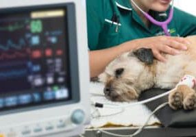
A multi-parameter monitor should be available to check SpO2, ECG and blood pressure. Ideally, we should obtain SpO2 result (in some cases this needs to be confirmed by a blood gas analysis of arterial blood to check PaO2, SaO2 and paCO2 levels). An approximation of etCO2 levels can be obtained by placing the end of the tube close to the nostrils of a nose-breathing dog and cat (avoiding obstruction of the nares or stress to the animal). This is especially achievable in recumbent animals.
Oxygen should be available in recovery and must be set up before moving the patient. Oxygen can be provided via flow-by method, mask, nasal prongs or in an oxygen cage. Recently the use of high flow oxygen therapy has been investigated in normal dogs (Jagodich et al., 2019) and dogs with respiratory disease (Pouzot-Nevoret et al., 2019) showing promising results. This technique requires specialised equipment.
References:
Brodbelt, D.C., Blissitt, K.J., Hammond, R.A., Neath, P.J., Young, L.E., Pfeiffer, D.U. and Wood, J.L., 2008. The risk of death: the confidential enquiry into perioperative small animal fatalities. Veterinary anaesthesia and analgesia, 35(5), pp.365-373.
Davis, H., Jensen, T., Johnson, A., Knowles, P., Meyer, R., Rucinsky, R. and Shafford, H., 2013. 2013 AAHA/AAFP fluid therapy guidelines for dogs and cats. Journal of the American Animal Hospital Association, 49(3), pp.149-159.
Jagodich, T.A., Bersenas, A.M., Bateman, S.W. and Kerr, C.L., 2019. Comparison of high flow nasal cannula oxygen administration to traditional nasal cannula oxygen therapy in healthy dogs. Journal of Veterinary Emergency and Critical Care, 29(3), pp.246-255.
Neamu, R.F. and Martin, G.S., 2013. Fluid management in acute respiratory distress syndrome. Current opinion in critical care, 19(1), pp.24-30.
Pouzot-Nevoret, C., Hocine, L., Nègre, J., Goy-Thollot, I., Barthélemy, A., Boselli, E., Bonnet, J.M. and Allaouchiche, B., 2019. Prospective pilot study for evaluation of high-flow oxygen therapy in dyspnoeic dogs: the HOT-DOG study. Journal of Small Animal Practice, 60(11), pp.656-662.
Case Advice or Arranging a Referral
If you are a veterinary professional and would like to discuss a case with one of our team, or require pre-referral advice about a patient, please call 01883 741449. Alternatively, to refer a case, please use the online referral form
About The Discipline
Anaesthesia

Need case advice or have any questions?
If you have any questions or would like advice on a case please call our dedicated vet line on 01883 741449 and ask to speak to one of our Anaesthesia team.
Advice is freely available, even if the case cannot be referred.
Anaesthesia and Analgesia Team
Our Anaesthesia and Analgesia Team offer a caring, multi-disciplinary approach to all medical and surgical conditions.