Please, please, please do not consider spontaneous non-traumatic haemoabdomen in the dog to be a surgical emergency. Despite some opinion leaders saying otherwise, our message to you is that emergency general anaesthesia and surgery are the two most assured strategies to accelerate your patient’s demise.
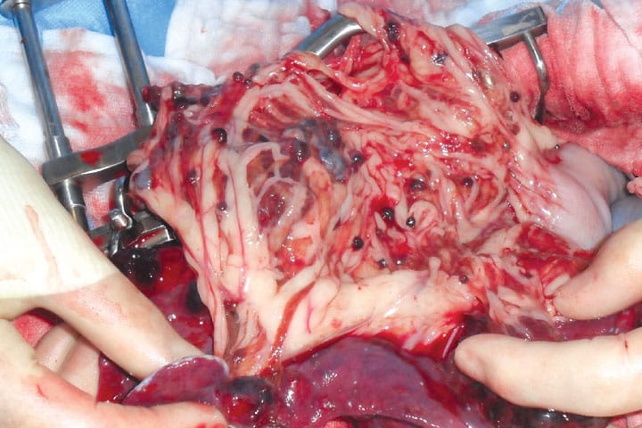
Spontaneous non-traumatic haemoabdomen in the dog does not only arise due to splenic haemangiosarcoma, but it is substantially the most likely cause. Most authors cite a 70-75% prevalence of haemangiosarcoma. In one study, 91% of cases proved to be due to haemangiosarcoma. Other reported causes include splenic lymphoma, splenic haematoma and hepatic abscess.

Pathophysiology
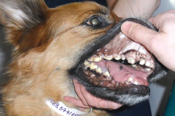
Patients are consistently hypovolaemic at presentation. A proportion of the circulating blood volume is free within the abdomen and not in their blood vessels. This leads to impaired blood distribution to vital structures. Despite the fact that whole blood is lost into the abdomen, these patients are usually also anaemic. This reflects the important fact that these patients have bled before. By the same token, there is usually thrombocytopenia. Platelets and coagulation factors are consumed by previous unseen bleeding episodes, impairing the patient’s ability to respond to further haemorrhage. When coagulation tests are performed using sensitive thromboelastography techniques, 100% of cases with splenic haemangiosarcoma exhibit a coagulopathy!
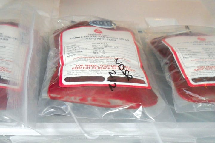
The emergency priority in cases with spontaneous non-traumatic haemoabdomen is correction of the physiological disturbances that have arisen due to hypovolaemia and reduced oxygen carrying capacity. Therefore, what these patients need more than anything, is red blood cells and fluid. These can be delivered as a fresh whole blood transfusion, or as the combination of packed red cells and isotonic electrolyte solution.

Differential Diagnosis
Unfortunately, it is very rare that we can be certain about the nature of the underlying disease process without histology. This means that clinicians will frequently have to decide whether to perform a potentially dangerous surgery without a diagnosis. There are certain clues available in the history, signalment or the further diagnostic investigations that can point you towards or away from a diagnosis of haemangiosarcoma (see table).

Surgery following recovery?
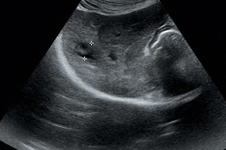
When normal circulatory function has been restored and the patient has become stable once again, attention should then turn to the question of whether to perform splenectomy. For most cases, the cause of bleeding was a splenic lesion but that is not always the case. If not already performed, thoracic radiography and abdominal ultrasound are advised as a matter of priority. Metastasis to liver and lungs is common and would be a contraindication to surgery. Splenic ultrasound would be expected to reveal multiple large, predominantly hypoechoic mass lesions. Findings to the contrary should prompt reconsideration of the wisdom of splenectomy. Of course, the spleen is a site of extramedullary haematopoiesis; removal could, in some cases, impair your patient’s recovery.

Histology
Have you ever received a histological report that declares your bleeding splenic mass to have been nothing more than a haematoma? It’s impossible to be happy about these because there is always the nagging doubt that in fact the mass was a tumour but that the tumour was simply not seen. It is definitely true that the pathologist has a much higher chance of returning an accurate diagnosis when the whole spleen is submitted for analysis. We recommend use of large sealable buckets for large specimens. The simple fact that the whole spleen cannot fit into a tiny universal container should not be allowed to prevent accurate diagnosis.
Medical therapy
In dogs with haemangiosarcoma, average life expectancy following splenectomy is consistently reported to be 1-2 months. This can be improved by the administration of chemotherapy. Broadly speaking, there are two chemotherapy strategies employed, conventional chemotherapy (e.g. doxorubicin) and metronomic chemotherapy (e.g. low daily dose cyclophosphamide). To date, the statistics favour conventional chemotherapy over metronomic chemotherapy. However, there are plenty of cases who are poor candidates for intravenous high-dose therapy for whom the attractive alternative of oral low-dose therapy is appealing. When asked what the ‘best’ treatment for splenic haemangiosarcoma is (following surgical removal) we suggest the combination of conventional chemotherapy first followed by metronomic chemotherapy once the initial course is complete.
Key messages
- Most dogs with spontaneous haemoabdomen have splenic haemangiosarcoma
- Emergency therapy should be directed at correcting the physiological disturbances of hypovolaemia and reduced oxygen carrying capacity
- In dogs with haemangiosarcoma:
- patients are hypovolaemic
- there has been chronic subclinical haemorrhage for a prolonged period
- there is coagulopathy in 100% of cases
- Clinical stage before surgery
- Histology can fail to identify tumours when only part of the specimen is submitted
Case Advice or Arranging a Referral
If you are a veterinary professional and would like to discuss a case with one of our team, or require pre-referral advice about a patient, please call 01883 741449. Alternatively, to refer a case, please use the online referral form
About The Discipline
Oncology

Need case advice or have any questions?
If you have any questions or would like advice on a case please call our dedicated vet line on 01883 741449 and ask to speak to one of our Oncology team.
Advice is freely available, even if the case cannot be referred.
Oncology Team
Our Oncology Team offer a caring, multi-disciplinary approach to all medical and surgical conditions.